Green fluorescent protein (GFP) is a kind of bioluminescent protein, found in coelenterates. GFP is easy to detect, high sensitivity, stable fluorescence properties, strong tolerance, easy to express, without cytotoxicity, used for living cell detection. Therefore, GFP can be used as a reporter gene to detect gene expression or regulation, or as a fusion label to detect the localization, migration, conformation change and intermolecular interaction of protein molecule, or targeted marker some organelles. The maximum peak of GFP absorption spectrum is 395nm (UV), and with a 470nm deputy absorption peak (blue ray). The maximum peak of emission spectrum is 509nm (green ray), and with a 540nm side peak (Shouder). 450nm-490nm only is the deputy absorption peak of GFP, but because the excitation light does less damage to cells, the band is usually used more frequently (mainly 488nm). In addition, the spectrum properties of GFP are similar to Fluorescein Isothiocyanate (FITC), both of two usually have a set of filters. GFP fluorescence is extremely stable. Under the excitation light, the ability of GFP Photo-Bleaching is stronger than fluorescein. In particular, it is more stable at the the blue ray wave of 450nm-490nm. Similarly, the sensitivity of the fluorescence of GFP fusion protein is higher than the fluorescent antibody of fluorochrome labeling, is strong anti-light bleaching ability, which is more suitable for quantitative determination and analysis. The generation of GFP fluorescence do not need to any exogenous reaction substrate. Therefore, GFP as a widely applied living reporter protein, its effect is unmatched by any other enzyme reporter protein. However, GFP is not enzyme. The fluorescence signal has no enzymatic amplification effect. So, the sensitivity of GFP is may lower than other enzyme reporter protein, like firefly luciferase.
The Ways of observing GFP:
1.Portable GFP Excitation Light Source
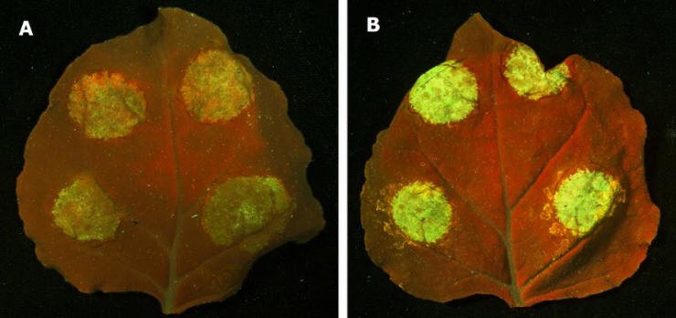
Using Portable GFP Excitation Light Source is the most convenient and fastest observation method (called Fluorescent Protein Observation Mirror). Because GFP has a higher absorption peak on 470nm, using 470nm excitation light source can stimulate GFP to emit bright green fluorescence. We must wear professional observation glass to observe the clear fluorescence signal when we observe GFP fluorescence. The reason is excitation light is intense blue light.
 |
| www.leduvcuring.com |
2.UV Light Source
Using UV Light Source ( called UV Light Lamp, Black Light Lamp) Observation is also very convenient. But if you observe the plant, leafy green of plant foliage also fluoresces under ultraviolet light. It is going to interfere for GFP fluorescence. Furthermore, the specimens may also cause damage to the GFP specimens under UV light.
 |
| https://www.leduvcuring.com/ |
3.Fluorescence Microscope
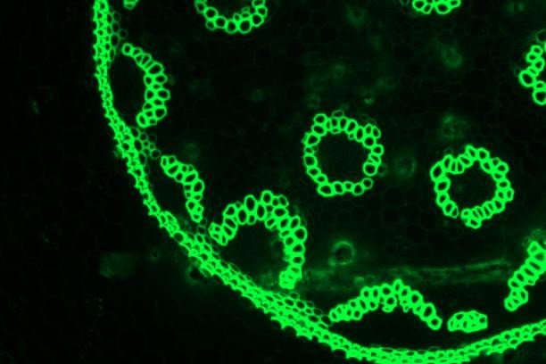
Fluorescence Microscope is based on UV as light source, is used to irradiate the detected object, makes it fluoresce, then observes the shape and location of the object under a microscope. Fluorescence Microscope is used to research the absorption of intracellular substances, transportation, the distribution and location of chemical substances. Some substances in the cell, like leafy green, can fluoresce after irradiation of ultraviolet light. Other substances, do not fluoresce in themselves, but if you dye it with a fluorescent dye or a fluorescent antibody, can also fluoresce by ultraviolet light. Fluorescence Microscope is one of the tools for qualitative and quantitative research on such substances. At present, the popular method is using Fluorescence Microscope observation. Fluorescence Microscope is very popular in laboratory. But the only deficiency is that the slices of leaf or animal must be collected for observation, which will destroy the cultured specimens and can not observe viviperception.
 |
| The reaction of GFP under fluorescence microscope |
Comprehensive Analysis, it is the most quickly and convenient method to observe GFP by adopting portable GFP excitation light source (called Transgenic Bios-Cope).
No comments:
Post a Comment